
New Technology May Help Inform Brain Stimulation
Summary: A new method that works by using ultrafast fMRI is in a position to capture brain exercise at sub-second amounts. The approach allows for serious-time checking of the brain underneath stimulation disorders.
Source: University of Queensland
Mind stimulation, this sort of as Deep mind stimulation (DBS), is a potent way to deal with neurological and psychiatric problems. When it has presented therapeutic gain for victims of Parkinson’s, Alzheimer’s, and addiction for more than a ten years, its underlying neural system is not however fully recognized.
Scientists at the Queensland Mind Institute (QBI) are now a single stage closer to unravelling the mystery of mind action to greater recognize this system and possibly predict DBS outcomes.
The brain is a very complex network of circuits organised hierarchically with broad-ranging connections. Connections go in unique instructions, forwards and backwards, and concerning neurons that are possibly excitatory – the accelerators of a reaction – or inhibitory – the brakes modifying a reaction.
“Say you want to shift your hand – when that signal is initiated, we count on that the activity that follows relies upon on the brain’s neural networks,” Associate Professor Kai-Hsiang Chuang said.
“What we really do not absolutely realize is how or when these structural and practical factors of the brain interact to inevitably lead to the end result of shifting your hand.”
Functional MRI (fMRI) is the most well known procedure utilised to examine mind networks. fMRI tracks blood flow and oxygenation variations following neural activity, thus indirectly measuring the functional connections being fashioned, and supplying us an sign of exactly where mind action is propagating.
Brain action, nevertheless, isn’t as simple as a signal travelling from location to space.
The workforce at the Chuang laboratory have designed a new ultrafast fMRI approach with a vastly greater temporal resolution, enabling them to seize the dynamics of brain activity at a sub-2nd amount.
Associate Professor Chuang reported the new approach had led to extra complete knowing of how and when the brain’s structural and functional connections interact.
“The first new discovery we designed is that mind action not only propagates as a result of structural wiring but follows specified preferential circuits depending on their excitatory and inhibitory neuronal distribution,” he explained.
“Communication concerning brain locations of related cell forms will become additional fluent, and the mind action much better.”
The Chuang group tracked the brain action of mice equally though stimulated and at relaxation making use of their ultrafast fMRI procedure. When the mind was stimulated, exercise adopted the structural wiring in the ahead route — from A to B and then B to C. When the mind was at relaxation, action was much more dependent on mobile form organisation and considerably less on structural wiring, propagating among C and B but not with A, if that’s wherever the preferential circuit was.
This indicates that how information and facts is processed is really dependent on your state, wherever it was previously believed that mind exercise functioned in the very same way irrespective of whether at rest or busy undertaking a task.
“The 2nd discovery we designed was that the blood signal detected by fMRI could reflect the community organisation and cell form distribution,” Affiliate Professor Chuang said.
“These conclusions have important implications for how brain structure shapes operate, and how to forecast action centered on the expertise of this construction. Additional pretty much, what we now know will affect the style of DBS and other brain stimulation techniques.
“The future techniques are to work with clinicians versed in brain stimulation to decide how we can utilise this understanding mixed with human information to help make improvements to our comprehension of DBS.”
This extra extensive being familiar with could empower us to superior predict DBS success and potentially increase its layout for superior therapeutic outcomes.
About this neurotech and DBS analysis information
Creator: Merrett Pye
Source: College of Queensland
Get hold of: Merrett Pye – University of Queensland
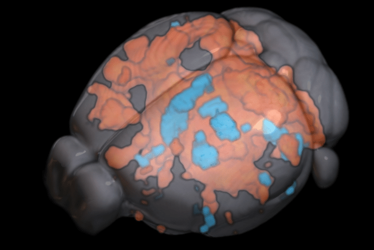
Image: The picture is credited to Associate Professor Kai-Hsiang Chuang / Queensland Brain Institute
Original Exploration: Shut obtain.
“Hemodynamic transient and functional connectivity observe structural connectivity and cell variety above the brain hierarchy” by Kai-Hsiang Chuang. PNAS
See also

Abstract
Hemodynamic transient and practical connectivity adhere to structural connectivity and mobile style about the mind hierarchy
The neural circuit of the brain is arranged as a hierarchy of practical models with extensive-ranging connections that support info stream and purposeful connectivity. Reports applying MRI suggest a moderate coupling involving structural and functional connectivity at the program stage.
Having said that, how do connections of diverse instructions (feedforward and feed-back) and areas with unique excitatory and inhibitory (E/I) neurons shape the hemodynamic action and practical connectivity over the hierarchy are unidentified.
Below, we used practical MRI to detect optogenetic-evoked and resting-state pursuits more than a somatosensory pathway in the mouse brain in relation to axonal projection and E/I distribution.
Working with a remarkably sensitive ultrafast imaging, we discovered in depth activation in locations up to the third order of axonal projections following optogenetic excitation of the ventral posteriomedial nucleus of the thalamus.
The evoked reaction and useful connectivity correlated with feedforward projections more than feedback projections and weakened with the hierarchy.
The hemodynamic reaction exhibited regional and hierarchical discrepancies, with slower and more variable responses in large-get areas and bipolar reaction predominantly in the contralateral cortex.
Electrophysiological recordings propose that these mirror distinctions in neural activity rather than neurovascular coupling. Importantly, the beneficial and unfavorable elements of the hemodynamic response correlated with E/I neuronal densities, respectively. In addition, resting-state functional connectivity was more related with E/I distribution, whereas stimulus-evoked efficient connectivity adopted structural wiring.
These results show that the structure–function romance is projection-, cell-type- and hierarchy-dependent. Hemodynamic transients could mirror E/I action and the increased complexity of hierarchical processing.